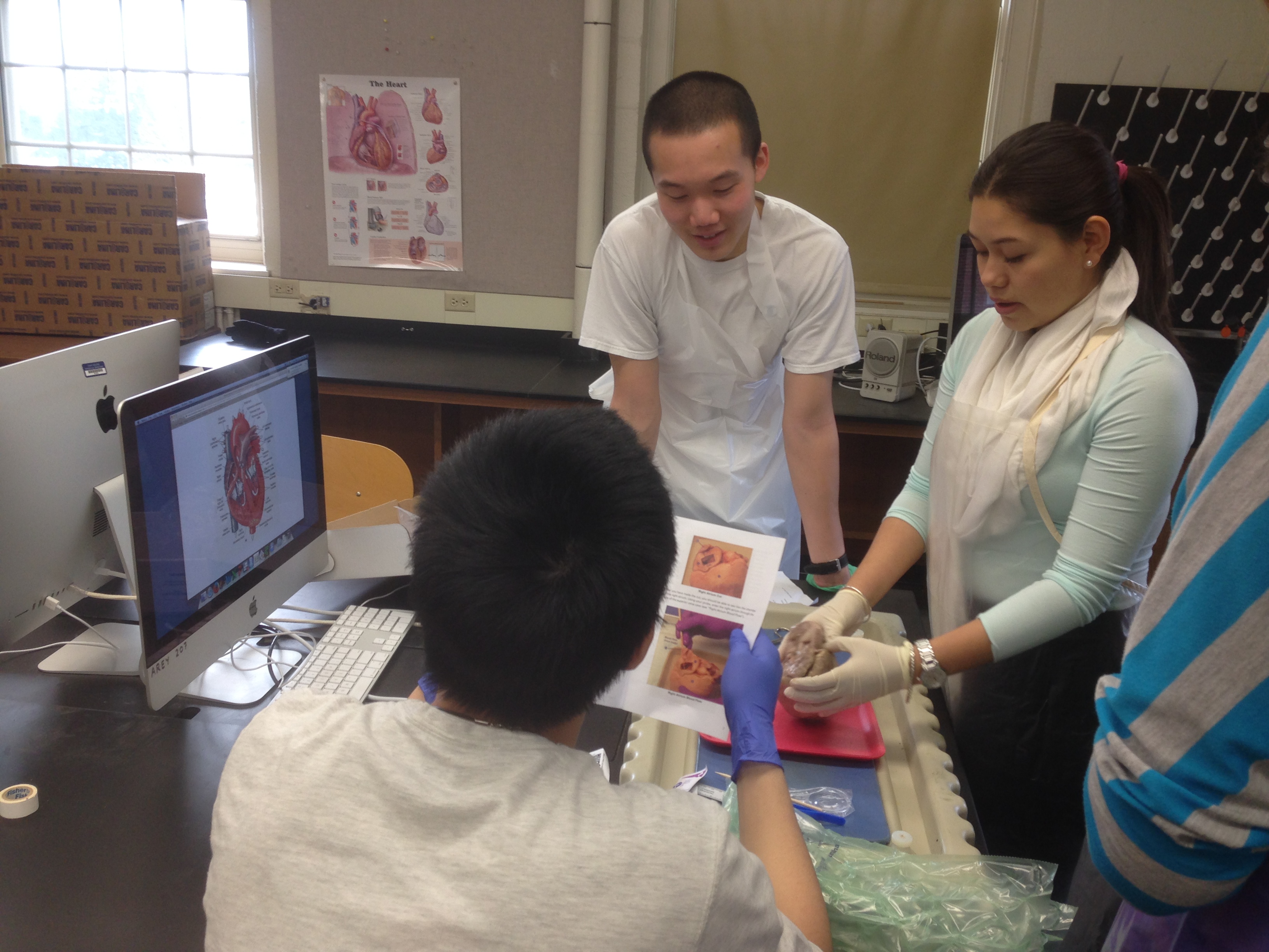
I cut the plastic bag open and was immediately hit with a strong odor. Inside the bag was a sheep’s heart, sitting in a pool of preservative chemicals. Immediately after taking the heart out, I went over to the sink to rinse off the chemicals. I could tell that this was going to be a messy lab. Still, I was excited to do a sheep heart dissection because it was an opportunity for me to see a real heart. For about a week, I had been studying diagrams and models of hearts, but not a real version of the organ itself. The anatomical models we used were helpful in learning where structures of the heart are located, but nothing beats seeing the real thing.
After washing the heart, my lab partner and I located its apex (tip) and figured out where the left and right sides were. It was immediately apparent that not all the structures were intact which was unfortunate (both the inferior and superior vena cava had been cut off). However, we discovered a well-preserved depression known as the foramen ovalis behind the right ventricle that had not been damaged in the preservation process of the heart. The foramen ovalis marks the former sport of the foramen ovale, a hole in the pig fetus’s that helps with blood circulation. After the pig’s birth, the hole is sealed, leaving behind the foramen ovalis.

Next, we started cutting the heart open. Cutting through the walls of the heart was difficult. The heart’s function, to pump blood throughout the body, requires it to be a tough, durable organ and I was reminded of this as my lab partner and I tried to cut through its walls. Pulling apart an incision on the right side revealed a small chamber with a hole in its lower end covered by three flaps. This was the tricuspid valve, the covering between the hole connecting the right ventricle and right atrium. I stuck my fingers through the valve, pushing my way past the three flaps into the larger space known as the right ventricle and felt around. Brushing against my fingers were the stringy cordae tendinae that connected the flaps of the tricuspid valves to the papillary muscles.
Over on the left side of the heart, we saw structures such as the left atrium, bicuspid valve, and the left ventricle. While making a cut on the left side of the heart, I immediately noticed how much thicker the muscular walls were on this side. Something that occurred to me during this dissection that I never really thought about before was how the heart’s form fit its function. Its thick, muscular walls (particularly on the left side) gave it the necessary power to pump blood to different parts of the body. The cordae tendinae, though somewhat thin and stringy, still felt strong and durable, which was necessary for them to be able to keep the bicuspid and tricuspid valves shut. Even the layout of the heart itself is essential to its function. It contains four chambers linked by a straightforward path that enables deoxygenated blood to enter, get pumped to the lungs to pick up oxygen, return, and then get pumped to other parts of the body. The sheep’s heart dissection was definitely my favorite activity from anatomy class. I enjoyed the hands-on aspect of it and being able to explore the layout and structure of a real heart.