Entries from January 2015
January 31st, 2015 · Comments Off on Danielle Levine: My Experience in Bi265j
Danielle Levine (’15, Biology)

To complete the Biology major at Colby, one has to take a minimum of six biology lab classes. As a senior biology major who at the end of the fall semester needed to take one more biology lab class, I chose to sign up for BI265 Introduction to Anatomy and Physiology for my January course rather than take an additional lab course (I will be taking the second semester of physics, which also has a lab) in the spring with my busy tennis team schedule. Having been warned before the class started that anatomy and physiology courses are a lot of work and a whole lot of memorization, I was expecting and prepared for an intensive month – but as the first week started, I found I was not quite ready for this class! During the first week, I was very nervous about the class – very concerned and stressed about the workload – and I remember wondering if I made the right decision to take the class, or if I should have just taken another lab course in the spring. After having now finished the class, I am very grateful for the opportunity to have taken BI265 with Dr. Klepach, as I truly enjoyed the class (excepting, of course, that first very difficult week!) to the fullest extent. I would recommend this class to every biology major, pre-med student, or any student at Colby just interested in learning how the human body works.
The class was, in fact, a lot of work, from studying for quizzes for almost every lecture, to listening to podcasts of lectures and labs for the next day, to making and presenting a Grand Rounds powerpoint to physicians and nurse practitioners, to studying for hours on end memorizing and identifying different anatomical structures and features on plastic models in the lab. However, the amount of material I learned and the understanding I achieved with respect to the structure and function of the human body was unimaginable to me before I took the class. The sheer volume of knowledge to be gained from this course is reason enough to recommend this class to other Colby students.
As a pre-med student, I am easily caught up with concern over my grades, GPA, exams and assignments, but taking this class reminded me of the importance of seeking to understand and learn material for oneself and for one’s own knowledge rather than for the primary purpose of getting a certain grade on an exam. As I hope to become a medical professional one day, this class had many practical aspects beyond the classroom; I was able to practice presentation skills via the class’s Grand Rounds presentation project, build a foundation of human anatomy and physiology for medical school (which I hope to go to!), learn how to succeed under stressful situations, and finally, balance my schedule keeping in mind the importance of maintaining a healthy lifestyle.
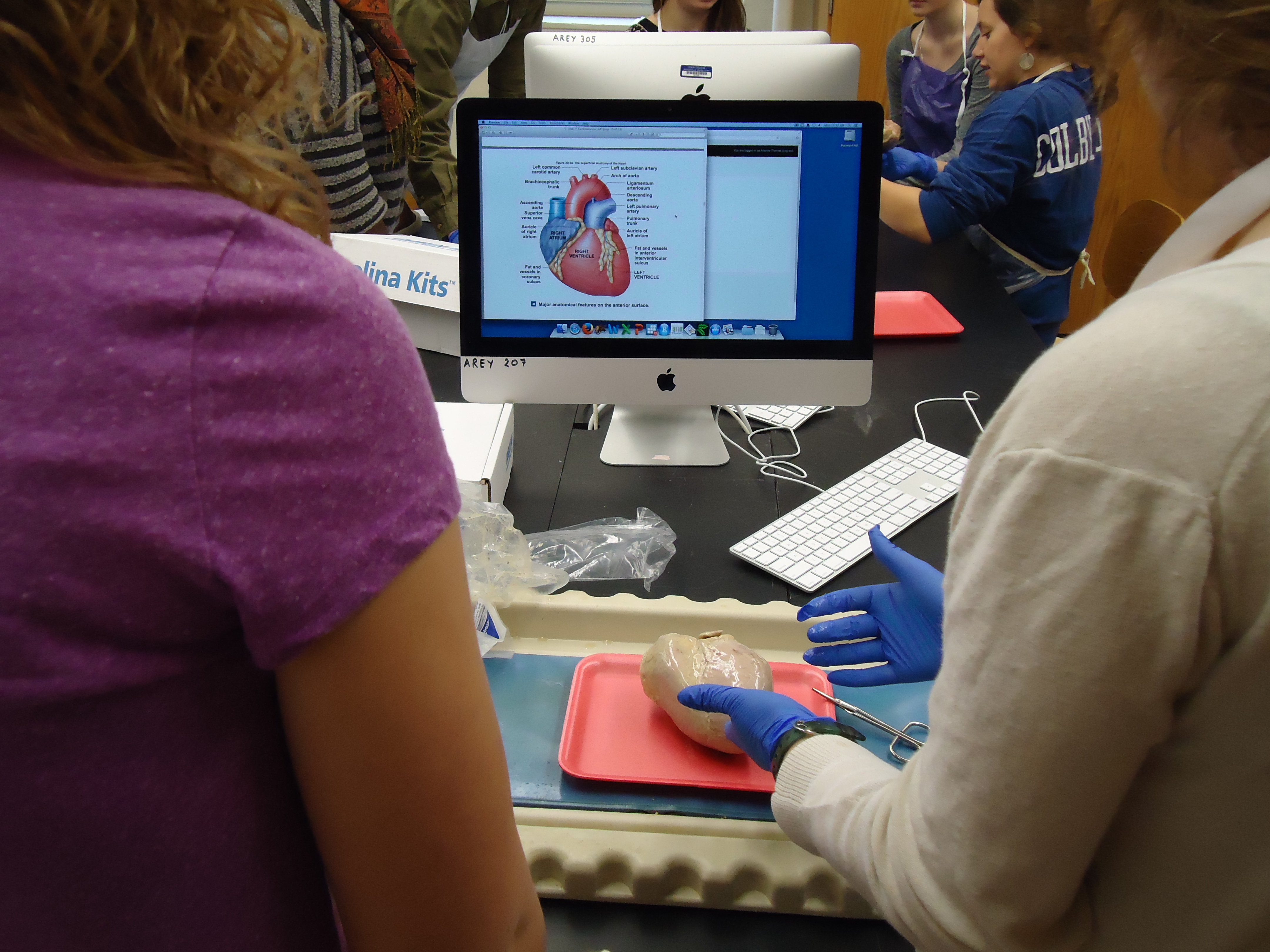
In taking this course, I was given many wonderful opportunities, such as being able to perform a wet dissection of a pig heart, and then being able to help visiting high school students perform a dissection on another pig heart, teaching them and sharing with them the material I had learned about the cardiovascular system the week prior, listening to guest lecturers, including Dr. Zak Nashed, who discussed interventional radiology and peripheral artery disease, and Dr. Peter Millard, who spoke about the field of epidemiology. Overall, I enjoyed this course very much, as it was a wonderful opportunity that I believe prepared me for the future after I graduate from Colby this spring, and reinforced my decision to pursue a career in medicine.


Here are some of the models used during lab and for studying for the lab practical exams.
Tags: Bi265j
January 31st, 2015 · Comments Off on Danielle Levine: Grand Rounds
Danielle Levine (’15, Biology)
One of the opportunities I had during this Jan Plan course was to participate in a Grand Rounds Presentation; Grand Rounds, in which physicians give lectures to their peers, including other physicians and medical students, on a medical topic is a common tradition in medical education. In groups of three students, we were able to pick any topic of interest for a fifteen minute oral presentation that we would present at the end of the semester. Given the vast array of medical topics that could be picked for a presentation, we looked to academic medical sources, including the New England Journal of Medicine, for possible past case studies that we could research and discuss. After scrolling through dozens of case studies, and clicking on articles with titles that seemed very interesting and then reading the articles’ summaries, we finally settled on an interesting case subject, one about celiac disease or gluten-induced enteropathy, that we considered particularly relevant given the current emphasis on the effects of gluten in the diet in popular culture.
In this case study, a 42-year old man presented to the emergency room with the chief complaint of chest and abdominal discomfort; given his additional history of unintentional weight loss and chronic diarrhea for ten years following coronary artery bypass grafting, an inflammatory disorder or a cancer of the chest or abdomen were differential diagnostic considerations. After multiple tests were performed, including an invasive exploratory laparotomy done after a CT scan showed enlarged jejunal lymph nodes, a small bowel biopsy revealed the diagnosis of celiac disease given the presence of flattened villi and intraepithelial lymphocytes. Today, celiac disease can be diagnosed via a simple blood test for IgA tissue transglutaminase and IgA endomysial antibodies. This case study demonstrates the importance for physicians, especially given the increasing incidence of celiac disease, to test for it non-invasively when a patient’s symptoms may be suggestive of it.
The diagnosis of celiac disease has been increasing in the developed world, at least in part due to the availability of new non-invasive tests to diagnose the autoimmune disorder. Also, there has been an increase in the diagnosis of non-celiac gluten sensitivity, which has been an even more significant factor in the increase in the number of people now adhering to a gluten free diet in the developed world. Unfortunately, some people do not have a true gluten-related disease or sensitivity, but are adopting a gluten free diet in a fad-like way. This is unfortunate because a gluten free diet can cause its own problems, such as nutritional (in particular, certain vitamins) deficiencies, and a lack of fiber in the diet leading to bowel-related issues.
Given the occurrence of a generalized increase in autoimmune disorders today, the hygiene hypothesis has been offered as a possible explanation, the basic tenet of which is that given increasingly prevalent strict hygienic practices, children today are exposed to fewer pathogens, and as a result can develop autoimmune disorders in which their own immune systems attack self antigens. Given the increasing numbers of people diagnosed with autoimmune disorders, it is hoped that research into celiac disease as well as other autoimmune disorders will lead to improved treatments of and ways to prevent them.
This Grand Rounds presentation was very informational as it allowed us to research a current topic of interest and, in so doing, learn the signs and symptoms that may exist at presentation of a certain disease (in this case, celiac disease), and how that disease may ultimately be diagnosed. As celiac disease is likely to continue to be a relatively common disease in the developed world, I believe this experience will be helpful to me, since I hope to become a physician someday. Being able to present our research to our peers as well as physicians and nurse practitioners, that is, to emulate something a real physician might engage in, was a wonderful experience. Furthermore, my mom, as a physician, talks about attending Grand Rounds Presentations every week at a hospital in New Jersey, and for me to be able to present a case study in the same manner was a fun and great opportunity.
http://youtu.be/DiKDOyG6Olg
Tags: Grand Rounds · Human Health
January 31st, 2015 · Comments Off on Cameron Matticks: JanPlan 2015 Internship talk to Bi265j Human A&P
Cameron Matticks, (’15, Cell & Molecular Biology) was the 2015 Bi265j TA and intern. Listen to his talk to the class about his experience below.
http://youtu.be/1uU5Tpgoaf8
Tags: Guest Speakers · Human Health · Internship Talks
January 30th, 2015 · Comments Off on Dr. Peter Millard, Epidemiologists Comes to Speak.
The last day of class we had the pleasure and honor of hosting epidemiologist Dr. Peter Millard, an MD PhD based in Belfast Maine, for a thoroughly engaging hour as he spoke about a wide range of epidemiological issues. The topics covered spanned his work on HIV infection in Africa, to the political, media and social components of disease right here in Maine.

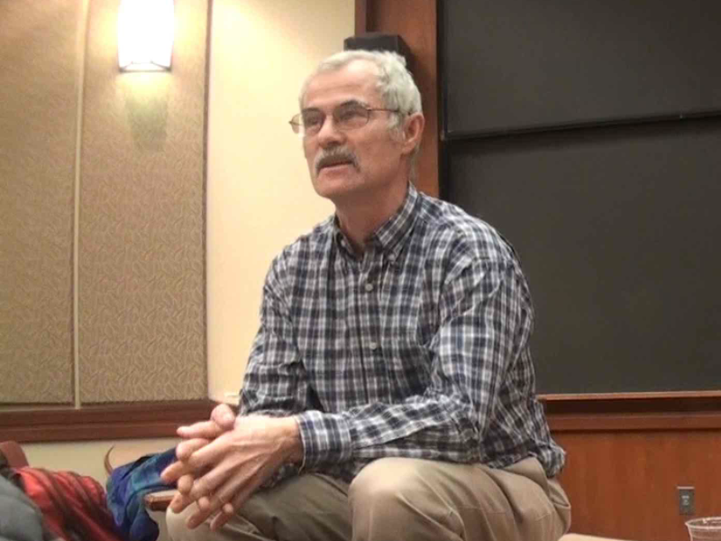

For all of the students in Bi265j I would like to thank Dr. Millard for graciously donating his time to come and speak to us. Watch his interesting and informative presentation below.
http://youtu.be/RFzfVgixaZU
Tags: Guest Speakers
January 30th, 2015 · Comments Off on Bi265j Mentioned in the Goldfarb Center Newsletter!
For the last two years the Goldfarb Center has generously supported a variety of activities in Bi265j Human Anatomy and Physiology through their Civic Engagement Course Development Grant. These activities include:
- A tour of Maine General Augusta and a presentation of their Grand Rounds talks along side similar talks from Human A&P students at Kents Hill prepatory HS to the third year UNE medical students on clinical rotations.
- Metabolic Analysis Lab in conjunction with the Waterville HS cross country team.
- Development of an internship for a former Bi265j student shadowing a nurse pratitioner in the Inland Hospital system in conjunction with being a TA for the Bi265j class.
- Bringing high school students interested in human A&P from a variety of regional school districts to campus for a day of mentoring by Bi265 students in collaboration with the Maine Math and Science Alliance. Activities included touring the anatomy lab, a hands-on pig heart dissection, and a brain storming session for helping the students develop human A&P based projects for the 2015 Maine State Science Fair.
These activities were recently mentioned in the latest Goldfarb Center newsletter which you can read here.
From myself and all of the Bi265j students at Colby and high school students from across the state that have benefited from the Goldfarb Center’s support, we give you our thanks. A special thanks to Alice Elliot, the Goldfarb Center’s Associate Director for her considerable logistical support and Amanda Cooley, the Assistant Director, for the write up in the GC newsletter.
Tags: Special Activities · Uncategorized
January 28th, 2015 · Comments Off on Grand Rounds: Atypical Hyperplasia of the Breast
Ariel Oppong, Jay Lee, Rebecca Gray
http://youtu.be/_0jV_CS7gXo
Grand Rounds _Hyperplasia powerpoint pdf
Grand Rounds Synopsis- Atypical Hyperplasia of the Breast
Hyperplasia occurs when an organ or tissue becomes enlarged because the cells within it begin to proliferate more quickly than usual, resulting in an abnormally large population of cells in one, concentrated area of the body. We categorize hyperplasia in two ways: (1) “simple” or “complex”, and (2) “usual” or “atypical”. The research we will explore focuses on complex, atypical hyperplasia. This refers to hyperplastic tissue that both engorges the tissue around it and contains deformed, non-uniform cells.
Hyperplasia of the breast falls into two categories: lobular and ductal. Within the fatty tissue of a healthy breast are mammary glands, and within those lie lobular clusters of alveoli. The cuboidal cells that line these alveoli secrete milk, which moves through milk ducts to reach the nipple, where it is excreted during breastfeeding. When hyperplasia occurs in the breast, it is usually found in either the lobules of the mammary gland or the associated milk ducts.
Usually, hyperplasia within the breast is relatively harmless. Because change in breast size and shape occurs normally over the course of a woman’s life, her body is designed to handle minor engorgement of the tissue there. This condition becomes concerning when hyperplastic cells within the breast become atypical; this is characterized by misshapen cytosol, nuclei, and membrane organization. When this occurs, the hyperplastic cells take on characteristics startlingly similar to those of tumors: they are clumped, proliferating rapidly, and lacking functionality. For this reason, atypical hyperplasia of the breast is linked with breast cancer and considered premalignant.
Usually if a lump or an abnormal mass is found during a female’s mammogram then a health professional will usually suggest a biopsy. During the biopsy tissue cells are removed for analysis by a pathologist. If the pathologist can not make a definite decision as to if the excision is cancerous or not.
A 2014 report published by the New England Journal of Medicine published a new meta-analysis of the associated risk of breast cancer associated with atypical hyperplasia. The conclusions of the meta-analysis suggested that a women with a atypical hyperplasia has a least a 30% increased risk of having breast cancer within a 25 year follow-up. Due to this new information we ( the anatomy group) as well as the authors of this newly published report agree that there needs to be a reform in women’s health policies so that women are more aware of their risk and are also more informed about some preventative medicine including screening techniques, and treatment or surgical options if signs of breast cancer are already apparent.
Some of the current commonly used treatments are different types of SERMS. SERMS are selective estrogen receptor modulators. The most widely used antiestrogen for management of breast cancer is Tamoxifen. However, prolonged use of Tamoxifen does increase one’s risk for endometrial (uterine) cancer.
Another important issue is the health disparities in breast cancer diagnosis, quality of treatment, and mortality rates among different socio-economic groups, geographic locations, the unemployed and employed, and racial groups. Based of off data from the 2014 Racial Disparity in Breast Cancer Mortality Study in areas such as Memphis, Tennessee, black women are more than two times more likely to die of breast cancer than their white counterparts. Overall, our research indicates that we as a country need to implement new screening methods, need to promote more education initiatives, need to enact new policies to decrease health disparities, and need to stress the powerful conclusions that meta-analysis provide.
Tags: Grand Rounds
January 28th, 2015 · Comments Off on Grand Rounds: Oligoastrocytoma
Grand Rounds: Oligoastrocytoma
Alex Lucas, Yvette Qu, Rachel Bird
http://youtu.be/wguUeUNttac
Grand Rounds_Oligoastrocytoma_powerpoint pdf
Oligoastrocytoma
Oligoastrocytomas are brain tumors that consist of oligodendrocytes and astrocytes, the two cell types in the brain that support and insulate nerve cells. Unlike many brain tumors, which typically present initially with headaches, seizures are a common initial symptom of oligoastrocytomas.
The patient presented with episodes of “a feeling of walking through a cloud,” receptive, or Wernicke’s aphasia (the inability to understand spoken words), Aphasia (inability to speak), and vertigo. She also suffered from brief seizures, which worsened in severity over the course of the next eight years, and began to involve loss of consciousness and muscle tone, occasional incontinence, and overwhelming confusion. Three months prior to admittance, the patient struck her head during an episode, but MRI, ECG, echocardiography, Holter monitoring, EEG and multiple blood tests all appeared normal. The patient did not respond to triptans or beta-blockers, but the frequency of her seizures increased to at least one per day.
An MRI was performed on the patient, which showed a mass in the left occipitotemporal region of the brain. A biopsy helped to determine the grade of the tumor – grade II, which is a low grade tumor – and also presents the pathology which helps to determine the growth patterns of the tumor cells. Fluorescence in situ hybridization (FISH), which allows for reliable and accurate detection of chromosomal deletions, showed deletions in 1p and 19q tumors.
Similar to most tumors, the exact cause of an oligoastrocytoma is unknown. It is understood that normal cells become abnormal in the sense that they may produce the wrong number of proteins or enzymes or be lacking certain genetic material. In the case of an oligoastrocytoma, deletions of genetic information in chromosomes 1p and 19q are the reason for the tumor cell’s abnormalities. This certain type of tumor is a result of a mixture of oligodendrocytes and astrocytes. Genetic material losses in 19q occur in 60-80% of oligodendrogliomas and 30-40% in astrocytomas, which demonstrate that there may be a shared variation in the formation of gliomas. Losses in the 1p chromosome are frequent with oligodendrogliomas at about 50-80%, however are less apparent with astrocytomas, which is detected only 10-18% of the time. However, the combination of genetic material losses of the 1p and 19q chromosomes is detected in 60-80% of oligoastrocytoma cases.
A gross resection was performed to remove the patient’s low-grade oligoastrocytoma tumor. Standard radiotherapy and antiepileptic medications were given after the resection. Lifelong MRI was suggested instead of permanent pacemaker due to the low-grade of the tumor. Following MRI shows only postsurgical changes, implying good prognosis. During the 24-month following the resection, no seizure has occurred with reduction in medication, indicating great possibility of freedom from seizure in 10 years and absence of intractability.
The patient’s case is complicated by ictal asystole (stopping of the heart during her epileptic seizures, which is very rare) in a patient with a predisposition to neurocardiogenic syncope (fainting, loss of consciousness, loss of muscle tone due to an abnormal control mechanism of the brain over the heart) due to a genetic disorder, and with the asystole being triggered by the seizures caused by her oligoastrocytoma make this case very interesting and unique. The important information the case conveys is that a patient’s symptoms are not always indications of a single disease, and sometimes the symptoms need to be closely examined and can suggest more than one disease. Perhaps screening for relationships between cardiac dysfunction and neurologic mechanisms could help identify rare cases such as this one, which would allow for earlier diagnosis and treatment.
References:
(1) Paleologos, N A, ed. Oligodendroglioma and Oligoastrocytoma. Am Br Tum Assoc 2014: 3-8.
(2) Oligoastrocytoma. Univ CO Sch of Med Neusrgy 2015.
(3) Meenakshi G, MD, Azita Djalilvand, MD, Daniel J. Brat, MD, PhD. Clarifying the Diffuse Gliomas. Am J Clin Pathol. 2005;124(5):755-768.
(4) Cole, AJ, M.D., Eskandar, E. M.D., Mela, T, M.D., Noebels, J.L. M.D., Ph.D., Gonzalez, R.G. M.D., Ph.D., McGuone, D, M.B., Ch.B. Case 18-2013 — A 32-Year-Old Woman with Recurrent Episodes of Altered Consciousness. N Engl J Med 2013; 368:2304-2312.
(5) Ucdenver.edu. Department of Neurosurgery [Internet]. 2015 [cited 2015 Jan 15]; Available from:http://www.ucdenver.edu/academics/colleges/medicalschool/departments/Neurosurgery/patientcare/multi-disciplinaryprograms/AdultBrainTumorProgram/Pages/Oligoastrocytoma.aspx
Tags: Grand Rounds
January 28th, 2015 · Comments Off on Grand Rounds: Postpartum Coronary Artery Dissection
Lauren Shirley, Allison O’Connor, Cal Robbins
Grand Rounds Synopsis
Case 28-2010 A 32-Year-Old, 3 Weeks Postpartum with Substernal Chest Pain
http://youtu.be/bODWv-x-eDc
Grand Rounds Case Presentation powerpoint pdf
Onset
A 32-year old woman had an uncomplicated, spontaneous vaginal delivery after 39 weeks of gestation. This was the patient’s second pregnancy. During her first pregnancy, she was diagnosed with preeclamptic toxemia which was treated with magnesium sulfate. Mild hypertension (systolic 120-140 mm Hg) was reported during the first and third trimesters of her second pregnancy followed by a return to normal blood pressure. Upon delivery, it was noted that her placenta weighed 340 g (below the fifth percentile for gestational age, mean 540 g) with increased amounts of perivillous fibrin (suggesting placental ischemia- lack of blood and thus oxygen and glucose to tissue).
The patient was admitted three weeks post partum when she developed pain in the left jaw and substernal area. The patient called EMS and was given oxygen which resolved her symptoms after 20 minutes and EMS personnel left. The pain returned shortly and EMS returned whereupon the pt scored her pain as a 7 out of 10. Blood pressure was noted as 148/74 and an electrocardiogram (ECG) revealed normal sinus rhythm of 90-100 bpm and ST-segment elevation of 4 mm in leads V2 and V3 (Abnormalities in ECG). Oxygen, acetyl-salicylic acid, nitroglycerin and morphine were administered. When examined at the hospital, the pt’s blood pressure was 143/92 mm Hg in her left arm and 137/81 mm Hg in her right arm with a pulse of 83-92 bpm.
Diagnosis
With a chief complaint of chest pain the patient could have been experiencing cardiovascular, pulmonary, gastrointestinal or musculoskeletal complications. Since the patient was 32 years old, cardiovascular complications would seem unlikely, however, since the patient was three weeks postpartum cardiovascular complications need to be considered more carefully since the risk of acute myocardial infarction is increased during pregnancy and the postpartum period and since pregnancy is a risk factor for aortic dissection. The risk of pulmonary embolism (a blockage of an artery in the lungs) is also increased during the postpartum period.
Since the patient’s ECG showed ST-segment elevation in conjunction with chest pain, an acute myocardial infarction would be suspected. Approximately 35% of postpartum women who present with myocardial infarction have a coronary artery dissection. There are two main types of coronary artery dissections, those that are caused by mechanical precipitation and those that are spontaneous. A spontaneous dissection is a tear in the artery where the tunica media and tunica externa separate, allowing blood to pool in between these layers. SCAD are rare, however 75% of patients who present with spontaneous aortic dissections are women and 30% of those women are peripartum, suggesting that this patient’s coronary artery dissection was spontaneous. There are four subgroups of spontaneous coronary artery dissections, however peripartum status and idiopathic spontaneous coronary-artery dissections or those caused by coronary shear stress are the two subgroups relevant to this case. Since the chest pain began after the patient picked up her toddler, there is a high index of suspicion that this dissection may have been caused by the patient’s peripartum status and coronary shear stress caused by lifting her toddler. Angiographic projections showed 35mm long segment of narrowing in the left anterior descending coronary artery. The lack of vascular disease in other coronary arteries along with the patient’s postpartum status as well as her test results are consistent with the diagnosis of a postpartum coronary-artery dissection.
Treatment Options
Unlike aortic dissections, the usual chest pain drugs (asprin, nitroglycerin, etc) which thin the blood can actually help, keeping the true lamen patent. Beta-blockers and nitrates are often used to prevent superimposed vapospasm. In cases of myocardial ischemia or compromised coronary flow, reperfusion therapy is used. In patients with severe ischemia, coronary-artery bypass grafting is done. In this case, the patient was given an intra aortic balloon pump which helps to increase myocardial oxygen supply by being placed in the aorta where it inflates and decreases based on the heart beat. Since the patient had no pain and the Percutaneous Coronary Intervention could have entered the false lumen, and since coronary dissections can heal by themselves, the balloon pump makes the most sense. This increased blood flow to the coronary artery. Aspirin as an antiplatelet, ß-blockers, and statins were used in case of intramural hematoma in the coronary vessel. Because of the potential for emergency cardiac surgery, the patient was not given glycoprotein IIb/IIIa inhibitors.
After 2 days a significant improvement was noted, the pump was terminated, and since surgery was now unlikely, glycoprotein inhibitors were initiated for a minor myocardial infarction discovered during treatment of the aortic dissection. This would be discontinued in a year, while aspirin was recommended indefinitely. The patient was able to return to her normal life with no further complications.
Little evidence exist in terms of the cause of spontaneous coronary artery dissections, but the current theory is that inflammation is caused by hormones, which explains the prevalence in post partum women. Several studies also included women taking oral contraceptives as being at risk for coronary artery dissections. The eosinophils release the histolytic agents between the tunica media and the tunica adventitia, which cause dissections in coronary arteries.
References
- Sabatine, Marc S., Farouc A. Jaffer, Paul N. Statts, and James R. Stone. “Case 28-2010: A 32-Year-Old Woman, 3 Weeks Post Partum, with Substernal Chest Pain.”The New England Journal of Medicine (2010): n. pag. Web.
- James, A. H. “Acute Myocardial Infarction in Pregnancy: A United States Population-Based Study.” Circulation 113.12 (2006): 1564-571. Web.
- Koul, Ashok K., Gerald Hollander, Norbert Moskovits, Robert Frankel, Leo Herrera, and Jacob Shani. “Coronary Artery Dissection during Pregnancy and the Postpartum Period: Two Case Reports and Review of Literature.” Catheterization and Cardiovascular Interventions 52.1 (2001): 88-94. Web.
- Mcintyre-Spatar, Leslie, and Kevin H. Silver. “Spontaneous Coronary Artery Dissection in a Postpartum Woman: Literature Review.” The Journal for Nurse Practitioners 7.9 (2011): 770-73.
- Oliveira Marta Silvia, Goncalves Alexandra, Dias Paula, Maciel Júlia Maria. “Spontaneous Coronary Artery Dissection: a Diagnosis to consider in Acute Coronary Artery Syndromes” Artigos de Revisão. (2009): 28 (6): 707-713
- Heart Assist Devices. Texas Heart Institute, 2015. (Accessed January 20, 2015 at http://www.texasheart.org/Research/Devices/iabp.cfm)
- CBC. MedlinePlus, 2015. (Accessed Janury 25, 2015 at http://www.nlm.nih.gov/medlineplus/ency/article/003642.htm)
- Placental Pathology. University of Chicago. (Accessed January 20, 2015 at https://pathology.uchicago.edu/sites/pathology.uchicago.edu/files/uploads/PDFs/Placental%20Pathology%20Notes%20Aspen%202014%20-Fritsch%20final.pdf)
- Electrolytes. AACC, 2013. (Accessed January 25, 2015 at http://labtestsonline.org/understanding/analytes/electrolytes/tab/test/)
- CK-MB. AACC, 2013. (Accessed January 25, 2015 at http://labtestsonline.org/understanding/analytes/ckmb/tab/sample/)
Tags: Grand Rounds
January 28th, 2015 · Comments Off on Grand Rounds: CABG v. PCI Stenting
CABG vs. Stenting in Multi-vessel Procedures: A Synopsis
Mayra Arroyo, Chris Lee, Ivan Yang
http://youtu.be/xsxlDjFb_sk
Coronary artery disease is caused by atherosclerosis, or the accumulation of fatty deposits, known as plaque, along the innermost layer of the coronary arteries. There are three main coronary arteries: the right coronary artery, circumflex artery, and the left anterior descending artery. Atherosclerosis causes the afflicted coronary artery’s wall to thicken and lose elasticity, ultimately narrowing or blocking the artery. This can reduce the oxygen flow to the myocardium. Treatment of coronary artery disease is complex and depends on several factors, but typically comprises of risk factor management, medication, and interventional techniques, such as coronary artery bypass grafting (CABG) and stenting.
Percutaneous Coronary Intervention (PCI) or Stenting is a minimally invasive process in which a doctor inflates a stent (mesh steel tube) with a balloon to open up a clogged artery. This restores normal blood flow. A catheter is inserted through the groin, neck, or arm to move the stent to the affected area. In recent years, newer types of stents such as drug-eluting stents and biodegradable ones have been developed. Before choosing stenting as a treatment option, one should consider risks involved with stenting such as damage to the vessels or arteries.
CABG is a surgical procedure where a vein or artery (usually from the inner thigh) is used to form a path around a blocked coronary artery. Over the years, there have been changes to the techniques used to carry out CABG. One such example is Totally Endoscopic CABG, which uses a robot equipped with a camera and surgical instruments in its arms. It is important to note that this procedure is highly invasive and risky, which is why it is usually used after more conservative treatments have been attempted.
In the three studies discussed, we compared long-term outcomes of CABG and stenting in multi-vessel disease in diabetics and in combined subgroups by looking at death rates, adverse event rates, and repeat revascularization rates. Multi-vessel disease is defined as the occlusion of two or more of the three main coronary arteries, and revascularization is a procedure that returns blood flow to a low-oxygen area.
In diabetics, we found that CABG ultimately has better long-term outcomes in multi-vessel treatment. Primary outcome (death, heart attack, or stroke) rates and all-cause mortality rates were lower in CABG diabetic patients than in diabetic patients who underwent drug-eluting stent procedures.
A study published in 2001 observed the effects that CABG and stent had on patients one year after treatment. The patients’ quality of life (survival and freedom from stroke, heart attack, or repeat revascularization) was examined after one year. It was found that there were no significant differences between the number of CABG and stent patients who did not die or have heart attacks or strokes. However, CABG patients had lower rates of repeat revascularizations than stent patients. In light of this study, it is important to remember that it was conducted in a time before drug-eluting stents and other newer treatment options.
Another study published in January of 2013 found that, compared to CABG patients, a greater percentage of people who underwent PCI with drug-eluting stents underwent repeat revascularization. The study concluded that in people with less complex disease, it is acceptable to undergo PCI. On the other hand, people with multi-vessel problems should choose CABG.
In conclusion, though CABG has proven to have better long-term outcomes than stenting in multi-vessel disease treatment, better studies should be conducted to verify this. Future studies should include more patients, cover modern CABG and stent techniques, and be extended for several years after revascularization. For now, it appears that CABG does have an advantage over stenting and drug-eluting stents, mostly due to reduced rates of repeat revascularization.
Tags: Grand Rounds
January 28th, 2015 · Comments Off on Grand Rounds: Celiac Disease
Ari Thomas, Laurel Edington and Danielle Levine
http://youtu.be/DiKDOyG6Olg
Grand_Rounds_Celiac_Disease powerpoint_pdf
Grand Rounds Synopsis
A 42-year-old man presented with a chief complaint of chest and abdominal discomfort that had begun suddenly two days before as a sharp left upper quadrant pain radiating to his back, associated with nausea and early satiety, and that increased in intensity over the next two days. The next day, he experienced substernal chest pressure consistent with his usual angina, but which did not respond to a single sublingual nitroglycerin tablet; it only resolved completely after IV morphine, ketorolac (an NSAID), chewable aspirin, and three more sublingual nitroglycerin tablets. He had experienced no recent abdominal trauma, vomiting, rectal bleeding or black stools.1
The patient’s past medical history includes hypertension, hyperlipidemia (excess blood lipids), and coronary artery disease (myocardial infarction at 32 years of age, with coronary artery angioplasty with stent placement, and subsequent bypass grafting).1,2 For more than ten years before presentation (since the coronary-artery bypass surgery), he has experienced chronic diarrhea that has worsened since his cholesterol-lowering medication was increased 6 months ago.1 During the past six months, he has experienced daily headaches, nocturia (excessive urination at night), feeling warm at night, an unintentional weight loss of 35 lbs, and occasional early satiety.1,2 Although he has a family history of colon cancer, a colonoscopy performed 4 months prior was unremarkable.1
The abdominal and chest pain, weight loss, and history of gastrointestinal symptoms suggested an acute chest syndrome, acute abdominal syndrome, inflammatory disorder, or cancer. An acute coronary syndrome was unlikely and an echocardiogram and chest radiography confirmed this. The physical exam ruled out acute chest syndromes, but sensitivity in the upper right quadrant suggested an upper abdominal disorder. Lipase, aminotransferase, and amylase levels were elevated, suggesting pancreatitis, liver injury (from an infection or drug use) or disease, or liver cancer. A CT scan of the abdomen ruled out pancreatitis and colon and small bowel disorders, but showed enlarged jejunal lymph nodes. An exploratory laparotomy was performed and the lymph nodes showed reactive follicular and interfollicular hyperplasia and lipogranulomas, suggesting an inflammatory abdominal disorder. Evidence of lymphoproliferative disorders was absent, ruling out cancer. A small-bowel biopsy showed flattened villi and intraepithelial lymphocytes, which suggested celiac disease. Positive IgA tissue transglutaminas and IgA endomysial antibody tests, the most specific and sensitive tests for celiac disease, were positive and confirmed the final diagnosis.1
The patient was advised to follow a gluten-free diet with an intramuscular iron supplementation as well as a multivitamin for general vitamin and mineral deficiencies.3 The gluten-free diet includes avoiding foods made out of wheat, rye, barley, oats, and processed foods that may contain wheat flour.4
Based on the patient’s symptoms, doctors diagnosed the patient with celiac disease. This disease is an autoimmune disorder that is provoked by intaking various forms of gluten and affects the small bowel. The intestinal symptoms of this disease include abdominal pain, diarrhea, a mild elevation of aminotransferase levels, and an increased risk of pancreatitis. Abdominal pain in the patient may have been due to transient intussusception related to celiac enteropathy. Celiac disease also has extragastrointestinal system effects such as rashes, arthralgias, neurologic and psychiatric effects, fatigue, and infertility.4 Patients also suffer from malabsorption of nutrients which can lead to weight loss, iron-deficiency, and osteoporosis. Patients have an abnormal immune response to the gliadin component of the gluten protein, where type 1 helper T cells cause inflammation in the epithelium and lamina propria of the small intestine, which alters the structures of the intestinal villi and therefore causes malabsorption.3 Celiac disease may also accompany type 1 diabetes, thyroiditis, and hepatitis.1
Celiac disease is different than a gluten sensitivity.5 Although the symptoms are similar, a person with a gluten sensitivity does not have the intestinal damage that a person with celiac disease has. Patients with a gluten sensitivity also do not have the IgA tissue transglutaminase or IgA endomysial antibodies that patients with celiac disease have.6 Since blood tests and intestinal biopsies will not diagnose a gluten sensitivity, using a process of exclusion helps to diagnose the sensitivity.5 Both disorders are treated by following a strict gluten-free diet.5,6
References:
- Ole-Petter Riksfjord Hamnvik, M.D., Fidencio Saldana, M.D., Bruce D. Levy, M.D., and Joseph Loscalzo, M.D., Ph.D. N Engl J Med 2014; 371:1333-1338.
- Medline Plus: Medical Dictionary. Besthesda, MD: U.S. National Library of Medicine, 2012. (Accessed January 13, 2015 at http://www.nlm.nih.gov/medlineplus/mplusdictionary.html.)
- Leffler, D. Celiac Disease Diagnosis and Management: A 46-Year-Old Woman With Anemia. Jama 2011; 306:1582–1592.
- Rubio-Tapia, A., Hill, I. D., Kelly, C. P., Calderwood, A. H., & Murray, J. A. American College of Gastroenterology Clinical Guideline: Diagnosis and Management of Celiac Disease. The American Journal of Gastroenterology 2013, 108:656–677.
- Non-Celiac Gluten Sensitivity. Ambler, PA.: National Foundation for Celiac Awareness, 2015. (Accessed January 25, at http://www.celiaccentral.org/non-celiac-gluten-sensitivity/).
- Gluten Sensitivity. Woodland Hills, CA.: Celiac Disease Foundation, 2015. (Accessed January 25, at http://celiac.org/celiac-disease/non-celiac-gluten-sensitivity/).
Tags: Grand Rounds
January 23rd, 2015 · Comments Off on Human A&P Grand Rounds Presentations

Our Human Anatomy and Physiology class will be presenting a series of talks on various diseases this coming Wendesday, January 28th 2015 from 9 until 11 AM on Colby’s campus in the Olin 01 auditorium, beneath the Olin Science Library. Each of the five 15 minute talks will be followed by a brief Q&A and will cover the following topics:
- Celiac Disease
- Oligoastrocytoma
- Atypical Hyperplasia of the Breast
- Postpartum Coronary Artery Dissection
- Coronary Artery Bypass Grafting vs. Stent Implantation
The presentation is free and open to the public and light refreshments will be served.
Tags: Grand Rounds · Human Health · Special Activities · Uncategorized
January 22nd, 2015 · 1 Comment
We were very lucky to have visitors to our class on Monday the 19th, MLK day, from a number of High Schools in Maine. The high schools included Lincoln Academy in Newcastle, Foxcroft Academy in Dover-Foxcroft, and even a home schooled Junior. The visit was designed to help the ten visiting students get a better sense of human anatomy and physiology in the hope of developing science fair projects for the Maine State Science Fair being held on March 21st in Bangor. The day was organized in conjunction with the Maine Math and Science Alliance and the Colby Goldfarb Center. For my part I was hoping to drive the material further into the brains of my students by following the aphorism the person who comes to teach learns the keenest lesson, while inspiring the spirit of mentorship towards the visiting students. The day started for my students at 9am with a practical lab exam covering the anatomy of:
- The central and peripheral nervous systems
- The eye and ear
- Sensory receptors
- The cardiovascular system and blood
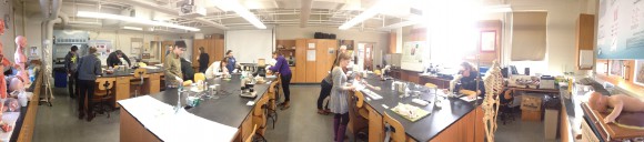

The lab has been up in Arey 307, typically the turf of microbiology lab, but for a month transformed into a splendid anatomy lab.

Danielle Levine (’15, Biology) contemplating a synaptic bouton during the test.

Lauren Shirley (’17, Biology/Music) looking at a dissected pig heart and Ariel Oppong (’16, Biology) inspecting an eosinophil in a histologic blood smear.

Mayra Arroyo (’16, Biochemistry) peering through a stereoscope at the optic chiasm on the 3D plate of a dissected brain from the Edinburgh Stereoscopic Atlas of Anatomy published in 1911.
Following the lab test the high school visitors turned up. I had initially intended for the visitors to start with an Art & Anatomy scavenger hunt similar to the one that I had designed for my students last week in the Colby Museum of Art, except this one would be based on clues created by the Colby students themselves, however the timing didn’t workout with the visitors being able to visit on their day off from school and MLK day falling on a Monday, the day that the museum is typically closed. Instead the students got to tour the lab and then participate in dissections of pig heart specimens. Rather than type out a description of the day I am simply going to reproduce the official event summary drafted by Stefany Burrell from MMSA, punctuated by annotated photos from the day taken by myself, Amanda Cooley of the Goldfarb Center and Stefany Burrell and Lynn Farrin of MMSA.
Notes from Colby J-Term Anatomy & Physiology Mentoring Session 1/19/2015
10:15 High school students from Lincoln Academy, Foxcroft Academy and a homeschool met Dr. Klepach’s class outside of the science buildings. It was a mild 40 degrees and sunny. Everyone headed into a lab in the Arey Building for an introduction.
10:30 Dr. Klepach welcomed the visitors and described his philosophy on science and teaching. The students were struck by his enthusiasm and knowledge. Many were inspired by his belief that teaching is learning.

Introduction by Stefany Burrell and Dr. Klepach.

Lynn Farrin (left) and Stefany Burrell (right) of the Maine Math and Science Alliance.
10:45 The students introduced themselves and the formed five groups, each with two high school students and three Colby students. Prior to this meeting, the class completed a lab exam. The exam consisted of approximately 30 questions in which students needed to identify various parts of human anatomy. The exam was broken into four sections: eyes, ears, nervous system and vascular system. As an icebreaker, the college students walked their charges through the exam, explaining what the physical models represent. The exam also included microscope slides, diagrams and a real pig hearts.

The Colby Human Anatomy and Physiology class started escorting their visitors around the test that they had finished less than an hour earlier.

Lauren Shirley is discussing the chambers of the heart with her fellow Colby students, Allison O’Connor (’17) and Cal Robbins (’17, Cellular/Molecular Biology) to the left and Dover-Foxcroft HS sophomores Bonnie (second from right) and Erika (far right).

Mayra, flanked by her Colby group members Ivan Yang (’17, Cellular/Molecular Biology, left) and Chris Lee (’17, Cellular/Molecular Biology, right) points to structures on a model of the heart to help Lincoln Academy seniors Abby (second from right) and Andrea (far right) understand what they are seeing on the dissected pig heart in front of them.

Erika getting a chance to look at the Edinburgh stereoscope slides.

Ivan discussing a cross sectional model of the spinal cord.

Can (John), a Lincoln Academy freshman (center), inspects a left coronary artery dissection as Colby students Yvette Qu (’18, left) and Alex Lucas (’17, Neuroscience & Sociology) look on.
11:10 The group moved to another lab where they put on gloves and aprons to dissect pig hearts. Each dissection station included a computer with loads of diagrams to assist in dissections. Under the Colby students’ guidance, the high schoolers dissected the hearts. Dr. Klepach moved around the room, answering questions as they came up. He took a few minutes to explain how blood moves into and out of the heart before and after birth.

Enormous cow heart ready for dissection.

Students preparing to dissect a pig heart try to orient themselves based upon surface anatomy.

Danielle discussing the surface anatomy of the heart with Cierra, a Dover-Foxcroft sophomore, and Shea-Lynn, a home schooled junior, as her classmates Ari Thomas (’16, Neuroscience, far left) and Laurel Edington (’15, Biology, second from left) look on.


Ashley (left) and Norma (center), seniors from Lincoln Academy, make the first cut into a pig heart as their Colby mentor, Rebecca Gray (’18, Biology / History), looks on.


Thilee, a senior from Lincoln Academy explores the left ventricle.

The aortic and mitral valves revealed!


Dr. K goes to the board to explain the flow of blood through the chambers of the heart.

12:00 Everyone got cleaned up and walked across campus to the Foss dining hall for lunch. Many people were drawn to the location as there was a noontime speaker in honor of Martin Luther King, Jr. Day. The crowd was thick and the supply of dishes and cups was low, but the food was delicious. Dr. Klepach had reserved a room for the group to eat lunch together. Many of the high school students were a bit overwhelmed trying to get their food amid such a crowd.

12:45 The next stop was the Olin Building, to a lecture hall below the science library. The students returned to their groups and Dr. Klepach introduced the final activity of the day: developing testable questions for science fair projects. Using a SMART Questions document produced by MMSA, the students came up with questions related to anatomy or physiology. They honed their questions and discussed how they might go about answering the questions.
One group had a good discussion about parameters that students can easily measure such as blood pressure, pulse, body mass index and body fat percentage.
Two other groups were curious about the physiological effects of various emotional states such as fear or amusement. They considered the use of video clips to trigger different emotions.
Another group, spurred by one student’s interest in livestock, was stumped by how they might measure parameters in a cow.
One pair of high school students, knowing that they would not be doing a science fair project, took the time to ask their mentor about college life.
The final group wanted to explore body image, comparing how people view their weight to reality. They came up with a good research plan that involved anonymous surveys asking people to describe if they think they are underweight, overweight or at a healthy weight. The subjects would guess at their weight and then use a scale to determine their actual weight.
Dr. Klepach asked each group to report out and asked thought-provoking questions such as how students would isolate variables. He also asked the students about the limitations of common measurements such as body mass index.

1:45 To wrap up the day, all participants filled out surveys. High school students and college students took separate surveys that asked about their motivations for participating, what skills they honed and what they considered to be the day’s highlights.
I thoroughly enjoyed having the visitors in the class and look forward to having them back in the future for this and other activities.
~Dr. K
Tags: Lab · Special Activities · Uncategorized
January 17th, 2015 · Comments Off on JanPlan 2015 Lab: Cranial Osteology, Art and Anatomy, and more…
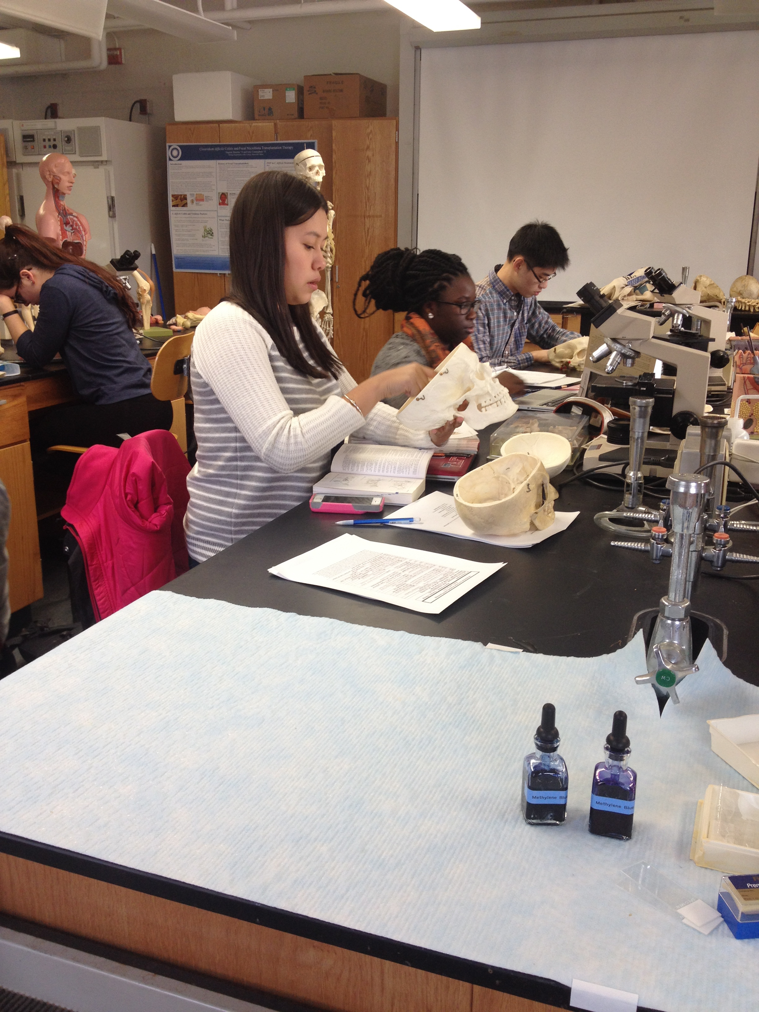
The active learner is the engaged learner, thus lab is an essential part of the semester here in Human Anatomy & Physiology. Here are a few images from lab so far…

Cameron Matticks (’15) was a student in the class in January 2014, and has returned as our TA this year as part of an internship that has had him shadowing nurse practitioners in the wound care unit at Inland Hospital in Waterville. Here he is sorting out the histology slides for an upcoming lab.

Left to right, Mayra Arroyo (’16), Ariel Oppong (’16), and Jay Lee (’15) in lab absorbed in the process of learning the osseous features of the cranium. Foramen magnum, foramen rotundum, foramen ovale, foramen spinosum… who knew there were so many holes in your head?

After the students learn the skeletal muscles in lab they get to test their knowledge in the Colby Museum of Art on the Art and Anatomy scavenger hunt. The students are given a specific muscle to search for that features prominently in an unnamed piece of art in the museum. As an additional clue they get a brief bit of art history on the piece in question. Here Ari Thomas (’16) contemplates John Rogers’ The Wounded Scout: A Friend in the Swamp as she searches for the flexor carpi ulnaris muscle.

Alex Lucas (’17) and Yvette Qu (’18) try to decide which clue to pursue next after finding the deltoidius muscle in Malvina Hoffman’s Bacchanale Russe. The sculptor was a master of human anatomy. Prior to the date of this particular casting, she had upon the advice of the famous Auguste Rodin, with whom she studied, traveled to the prestigious Columbia University College of Physicians and Surgeons in New York City, the town of her birth, to spend a year dissecting human cadavers and learning human anatomy, highlighting the close relationship between anatomy and art that has persisted throughout the ages.
Tags: Lab